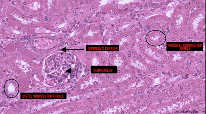
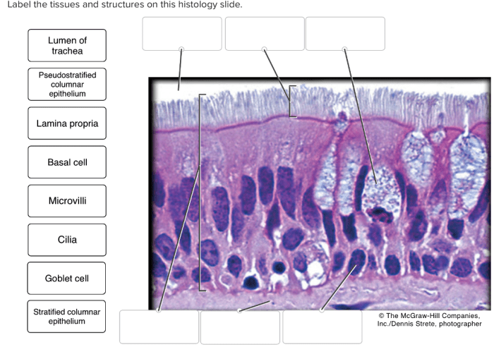
Label the the tissues and structures on the histology slide – Labeling histological tissues and structures is a fundamental step in histology, providing a systematic approach to identify and understand the various components within a tissue sample. This guide offers a comprehensive overview of the process, outlining key principles, techniques, and best practices for accurate and effective labeling.
By precisely identifying and describing the tissues and structures present in a histology slide, researchers and medical professionals gain valuable insights into the structure, function, and pathology of organs and systems. This information is crucial for diagnosis, treatment planning, and advancing our understanding of human biology and disease.
Label the Tissues and Structures on the Histology Slide

Labeling the tissues and structures on a histology slide is essential for understanding the cellular organization and function of the tissue. By accurately identifying and labeling the various components, we can gain insights into the normal histology of an organ or system and identify any abnormalities that may be present.
Identify the Various Tissues and Structures
- Identify the different types of tissues present, such as epithelium, connective tissue, muscle tissue, and nervous tissue.
- Identify specific structures within each tissue, such as epithelial cells, collagen fibers, muscle fibers, and neurons.
- Describe the morphology and key characteristics of each tissue or structure, including its shape, size, arrangement, and staining properties.
Organize the Information
To facilitate easy reference and understanding, organize the information in a table with columns for tissue/structure name, description, and location. Use bullet points to list the key features of each tissue or structure. Consider using color-coding or highlighting to emphasize important information.
Provide Detailed Descriptions
Explain the function and significance of each tissue or structure in the context of the histology slide. Discuss the relationships between different tissues and structures, and how they contribute to the overall function of the organ or system. Include relevant clinical correlations or pathological implications where appropriate.
Illustrate with Images, Label the the tissues and structures on the histology slide
Incorporate high-quality images of the histology slide to illustrate the labeled tissues and structures. Use arrows, labels, or overlays to clearly indicate the specific features being discussed. Provide captions or legends to explain the images and their relevance to the analysis.
Essential FAQs: Label The The Tissues And Structures On The Histology Slide
What is the purpose of labeling histological tissues and structures?
Labeling histological tissues and structures allows researchers and medical professionals to accurately identify and describe the various components within a tissue sample. This information is crucial for understanding the structure, function, and pathology of organs and systems.
What are the key principles of labeling histological tissues and structures?
Key principles include using precise anatomical terminology, organizing information in a logical manner, providing detailed descriptions, and illustrating with high-quality images. Accurate labeling ensures consistent and reliable analysis.
What are some common challenges in labeling histological tissues and structures?
Challenges may include distinguishing between similar structures, identifying rare or unusual tissues, and interpreting complex histological patterns. Careful observation, reference to anatomical atlases, and consultation with experts can help overcome these challenges.

